High-content screening (HCS) is an advanced form of high-throughput screening that leverages automated microscopy and live-cell image analysis to extract detailed cellular information.
This powerful technique enables the identification of compounds affecting cellular morphology, protein localization, and other features across vast compound libraries. HCS can be applied to various cellular systems, from individual cells to complex 3D cultures, without prior knowledge of specific therapeutic targets. At Assay.Works, we employ premier image acquisition and analysis capabilities for phenotypic assays, 3D cultures, and high-content screening. Our hit.works robotic liquid handling platform, equipped with two Operetta CLS™ image acquisition systems, allows us to establish complex physiological readouts and cell phenotypes with low variability and a daily throughput of up to 5 TB of data. This advanced setup enables efficient hit identification and subsequent target deconvolution using cutting-edge technologies like genome-wide siRNA analyses and CRISPR-based approaches.
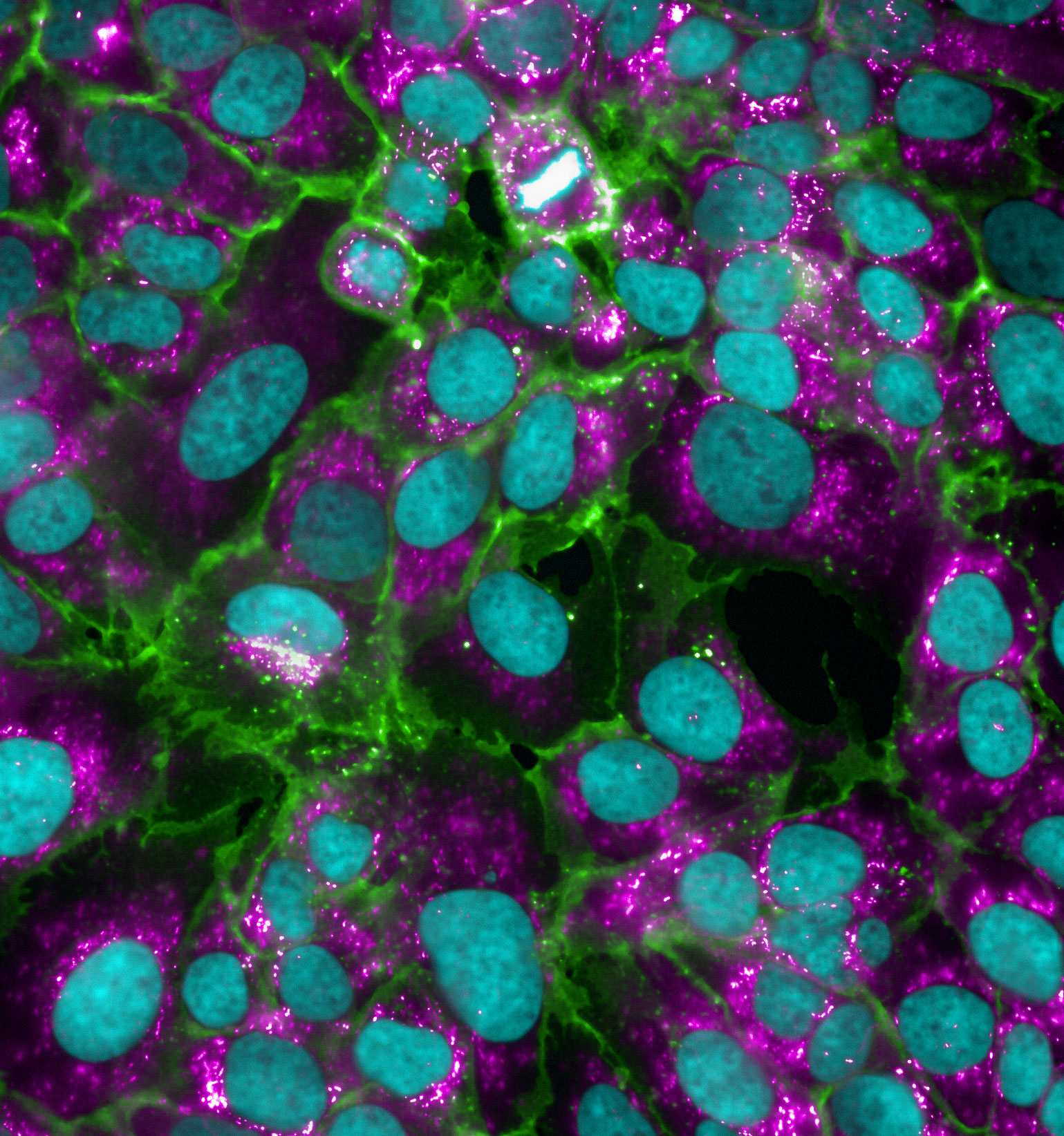
Cell Models & Readouts
- Engineered cell lines
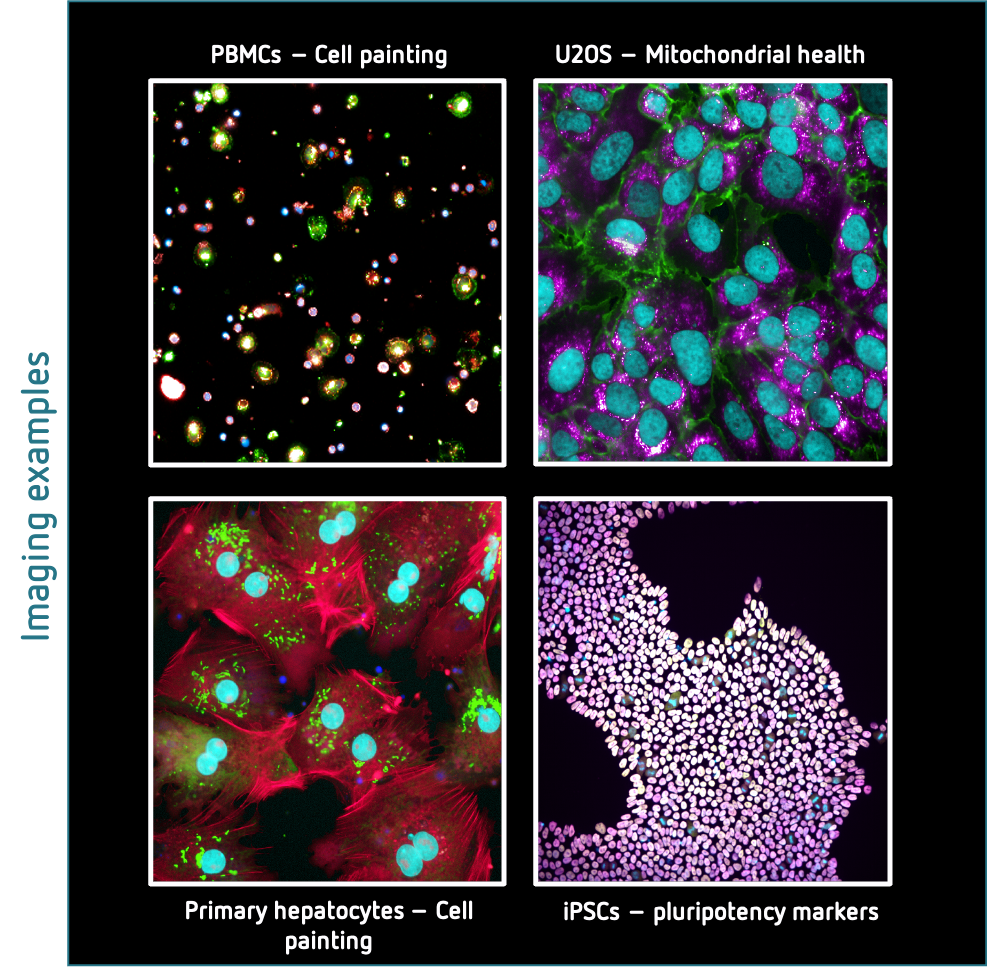
- Peripheral blood mononuclear cells (PBMC)
- Primary human hepatocytes
- Hepatic stellate cells
- Induced pluripotent stem cells (iPSC)
- Primary muscle cells
Imaging-based Readouts
- Cell Confluency (Brightfield, Digital Phase Contrast)
- Fixed cell staining (dyes, immunohistochemistry, CELL PAINTING panel)
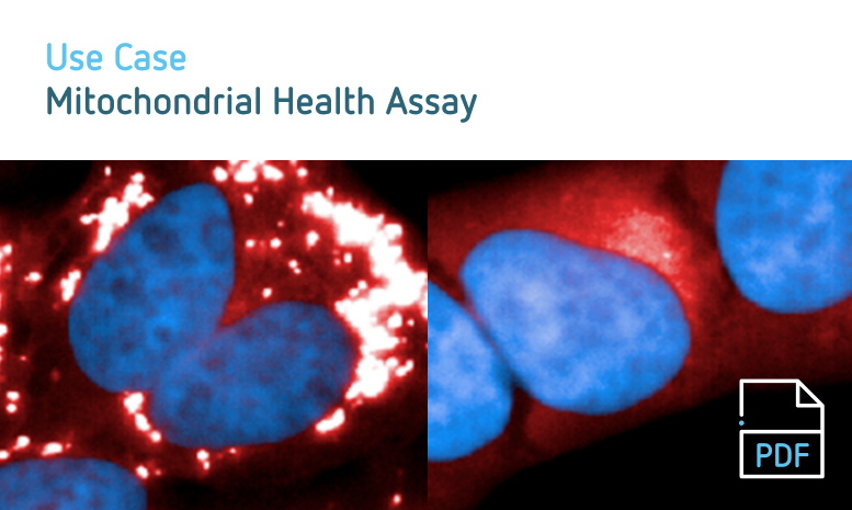
- Live-cell staining in temperature & CO2 controlled environment, e.g. mitochondrial markers, cell viability, membrane, or nuclear dyes
- Data Infrastructure
Custom Cloud Storage Upload Solutions
- Compatible with major providers:
- Amazon S3
- Google Cloud Storage (GCS)
- Microsoft Azure Blob Storage:
Tailored to specific organizational needs
On-Premise Storage Solution
- Server-grade storage system
- High capacity: 36 TB per Operetta
- Operetta throughput 5 TB images/day
Key Features
- Reliable, enterprise-level hardware
- Designed for instruments generating large data volume
- Scalable to accommodate future growth
- Compatible with major providers:
- Image Acquisition Workflow
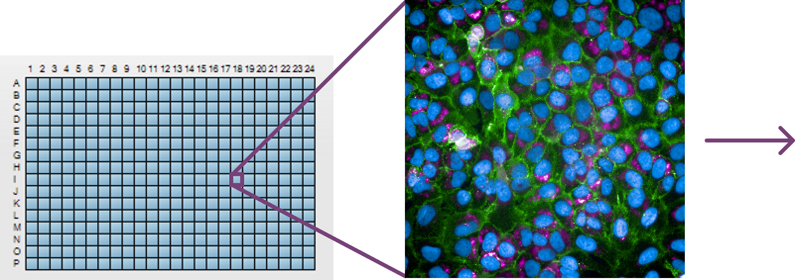
- Capture high-resolution microscopy images of prepared cellular samples on Operetta CLS
- Perform nucleus and cell segmentation to analyze on single-cell basis
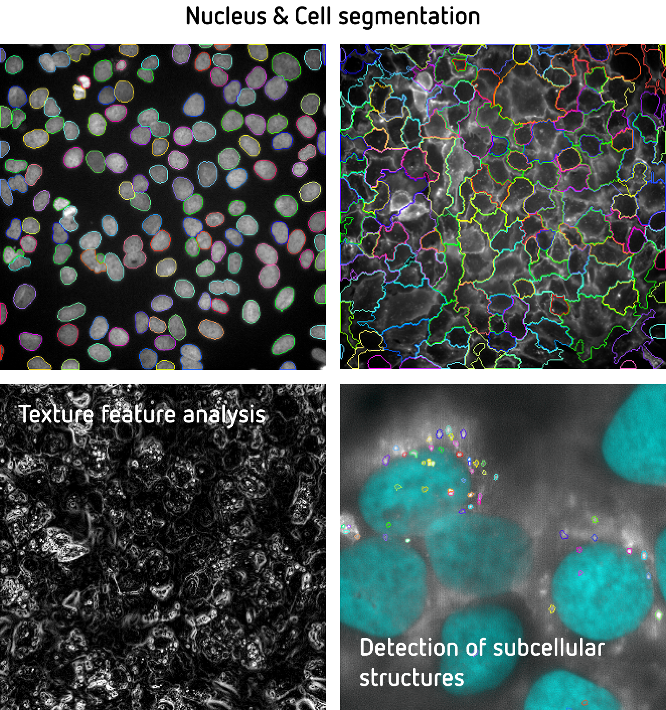
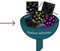
- Extract cellular features (morphology, texture, spatial etc.)
- Analyze data to identify patterns, classify cells, or detect morphological changes
- HCS Data analysis
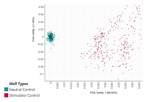
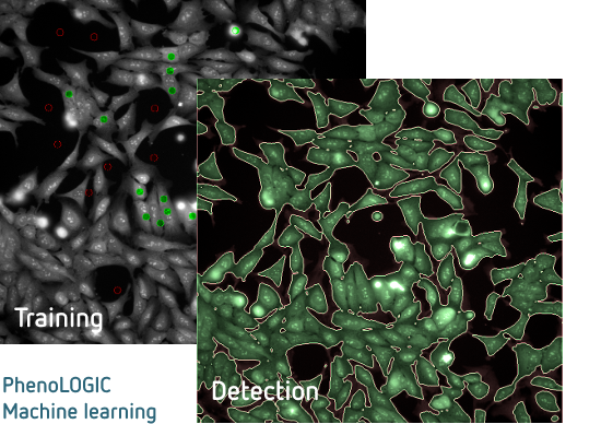
Assay.Works employs advanced data analysis techniques for HCS using Genedata Screener, which incorporates Principal Component Analysis (PCA). This powerful statistical method reduces dimensionality in complex imaging datasets, enabling researchers to identify key patterns and variables in cellular responses. PCA effectively reduces noise, compresses data while preserving crucial information, and enhances machine learning model performance for HCS. The software can score texture features like Haralick, Gabor, and SER using optimal data classifiers between controls. Additionally, Assay.Works utilizes Harmony Software, which offers tools for intricate cellular analysis, including 3D imaging and multi-parametric measurements. Harmony's PreciScan Intelligent Acquisition significantly reduces acquisition and analysis times, particularly beneficial for 3D microtissue and rare-event studies. The PhenoLOGIC feature automates complex phenotypic classifications, improving consistency in cell population identification through machine learning and minimizing the need for manual intervention in analysis workflows.
